ILD SPECIALIST

Dr. Sheetu Singh is a woman of dynamic personality with a noble heart. Comfort care and quality cure are the primary concern for her and this comes with trust that she gained from her clients. Being Ild Specialist in Jaipur Rajasthan India she has various equipments to cure this deadly disease.

What is ILD?

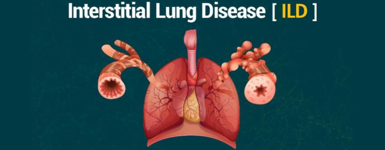
Interstitial lung disease (ILD) is a group of disease in which the lungs get shrunken due to multiple causes. The symptoms of ILD are cough without sputum and difficulty in breathing while walking or exercise.

What causes ILD?

ILD is of different types the common ones are: hypersensitivity pneumonitis (HP), connective tissue associated ILD, idiopathic pulmonary fibrosis (IPF) and sarcoidosis.
HP is the most common type of ILD and is caused due to exposure to birds such as pigeons, parrots, hen, molds on wall, leaky airconditioners and aircoolers with dirty old mats.
Connective tissue disease associated ILD is due to disease such as rheumatoid arthritis, scleroderma and sjogren’s syndrome.
The cause of IPF is unknown. However it is seen in elderly men who have history of smoking.
Sarcoidosis also the cause is not known. Though, its association with tuberculosis has been shown in studies.

What are the symptoms of ILD?

  • Difficulty in breathing: on walking, climbing stairs and exercise
  • Cough with minimal sputum: sometimes cough is very distressing
  • Chest pain
  • Joint pains – in patients with sarcoidosis and connective tissue associated ILD
  • Raynaud’s phenomenon – fingers turn blue on exposure to cold. This happens in patients with scleroderma associated ILD

How to diagnose ILD?

Chest x ray, spirometry and six minute walk test provide clues that patient may be having ILD. Confirmation is done with the help of special type of CT scan called as HRCT.

Further classification in various types of ILD requires a battery of tests which include

  • Serum collagen profile: helps differentiate connective tissue associated ILD
  • Serum IgG for birds: helps detect bird exposure in cases of HP
  • Bronchoscopy and transbronchial lung biopsy: may be planned to take a piece of lung for examination
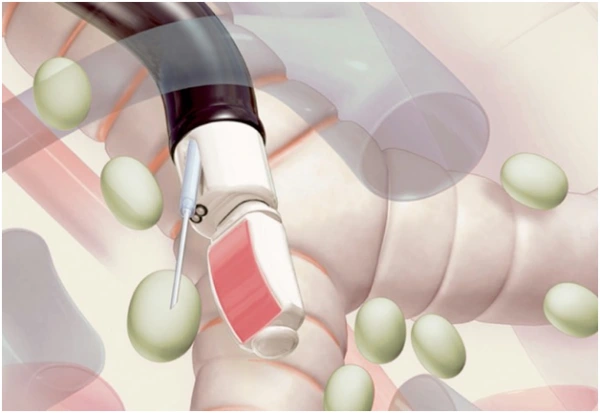
  • EBUS TBNA: this is a specialized type of bronchoscopy which takes samples from the lymph nodes in patients with sarcoidosis


ILD specialist in jaipur, Rajasthan, India

How to treat ILD?

There are 2 types of ILD –

  1. Steroid responsive: especially in cases of HP, connective tissue associated ILD and sarcoidosis. These diseases have good response to steroids. Certain steroid sparing agents may be used called as immunosuppressive agents. These help in bringing down the medication dose. Immunosuppresives drugs include azathioprine, mycophenolate mofetil and cyclophosphamide.
  2. Steroid unresponsive: IPF is a disease which does not respond to steroids. The recommended drugs are Pirfenidone and Nintedanib.

How to check treatment response?

Spirometry, diffusion study and six minute walk test are simple and easy test to check for treatment response. CT scan is repeated only after 1-2 years.

Cryo-biopsy for diagnosis of Interstitial lung disease (ILD)

Interstitial lung disease (ILD) is associated with significant morbidity and mortality. The diagnosis of ILD is largely based on clinical history, high resolution computed tomography (HRCT) chest and certain ancillary investigations. Despite these there is a subset of patients for whom the diagnosis is unclear. Traditionally, there were 2 types of biopsies that were being done including transbronchial lung biopsy and VATS associated surgical lung biopsy. Transbronchial lung biopsy is associated with smaller size, lower diagnostic yield and complications such as bleeding and infection. VATS associated lung biopsy is the gold standard but is associated with risk of infection, exacerbation of underlying ILD, persistent air leak, and prolonged ventilation. The need for less invasive and more safe procedures was there which led to evolution of cryo-lung biopsy. Investigations done prior to biopsy: routine blood tests would be done including complete blood counts, renal function test and bleeding profile. 2D echocardiography is done prior to rule out moderate to severe pulmonary hypertension. Spirometry may also be done. COVID-19 RT PCR is done as per the local prevalence of COVID-19 at the time of procedure. If the pre-operative evaluations were within normal limits then the patient is posted for the biopsy.

Procedure:
The procedure is done under general anesthesia. No incision is required. A endotracheal tube or rigid bronchoscope is used through which the flexible bronchoscope is inserted. An occlusion balloon is used to provide tamponade and control bleeding. 4-6 biopsies are taken under C-arm guidance to lower the risk of pneumothorax. Complications: bleeding controlled by occlusion balloons and risk of pneumothorax lowered by use of c-arm.

Post-operative course:
the patient is usually extubated on the table and is followed up for 24 hours. A chest X ray is done 4-6hours post procedure to rule out pneumothorax. The biopsy is reported by the pathologist overy 48 to 72 hours. It has to be discussed with your doctor to change treatment plans.


© 2017 Dr. Sheetu Singh. All rights reserved | Webworks by Theacemakers.com

Codeskube Pvt Ltd